In our previous two articles, Oral pathology of Oral Cavity and Oropharyngeal Squamous Cell Carcinoma, and Oral Pathology of Oral Pharyngeal Squamous Cell Carcinoma and HPV, we looked at histological images, discussed biological profiles, trends, survival rates, and treatments. In this article we will provide additional clinical pictures of Squamous Cell Carcinoma.
Don’t have time to read this article? We get it. Download the Diagnosing Vesicular Ulcerative Conditions checklist to get the key information and images from this article plus all the other conditions we cover in the Dentist’s Guide to Oral Pathology.
Clinical Pictures of Squamous Cell Carcinoma Affecting the Gingiva
Here’s a large ulcerative granulomatous type lesion. This was a squamous cell carcinoma of the gingiva.
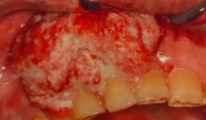
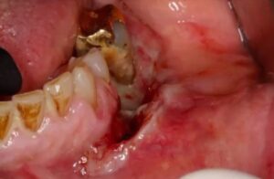
Here’s a more ulcerative, granulomatous example. This patient had an extraction and the clinician didn’t think to biopsy the area and thought that this extraction socket just wasn’t healing properly. This granulation tissue turned out to be a cancer.
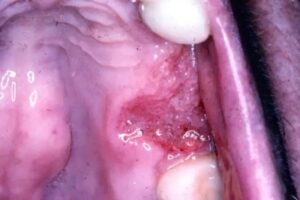
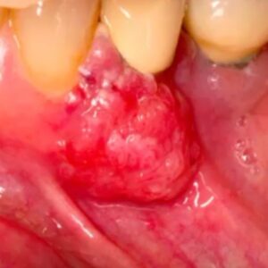
Here is a squamous cell carcinoma that closely mimicks pyogenic granuloma. Because there’s a lot of calculus here you might think that it is pyogenic granuloma, but this is abnormal and needs to be biopsied. Never take tissue and throw it away. Always submit it for biopsy.
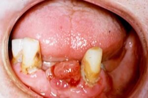
Same thing happening here. Generally, the dentist’s were working on teeth in the area and thought for several months that it was just granulation tissue. In these patients, two separate patients, both of these turned out to be squamous cell carcinomas.
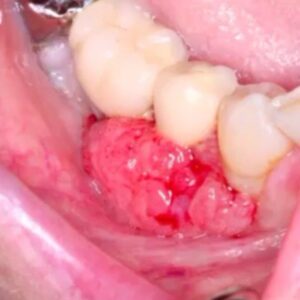
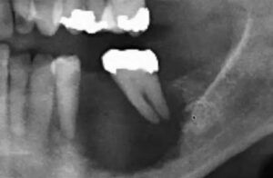
Here’s an obvious squamous cell carcinoma involving the gingiva with the corresponding radiograph. It’s never good when you’re seeing a tooth floating in air. You’re seeing a regular, poorly defined periphery of this lesion which is not a good sign.
Clinical Pictures of Squamous Cell Carcinoma with Periodontal Involvement
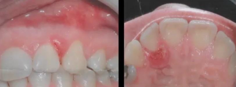
The dentist was working on a dental implant. They noticed that the patient has good oral hygiene so they wondered why only one tooth was periodontally involved and needed to be extracted. At that point in time there was cancer already. The cancer spread all the way here. This is a major concern. If you see something abnormal, don’t ignore it. Take a biopsy put it in formalin and send it to a lab.
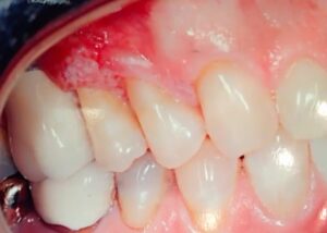
Sometimes squamous cell carcinomas are less obvious. This patient, a young female, had very good oral hygiene. Why does this patient have something that looks like a pyogenic granuloma when she’s got very good oral hygiene? This is the lingual view and again ulcerated lesion. They took a biopsy and turned out to be a squamous cell.
Periodontitis and Squamous Cell Carcinoma Pictures
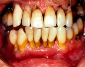
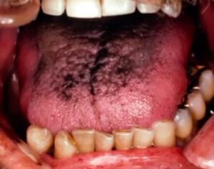
Here’s an example of periodontitis. If you are thrown off by the periodontal disease and the black hairy tongue and you don’t do your oral exam properly you’re gonna miss the squamous cell carcinoma. This patient came in for a cleaning and had no idea that this lesion was there. Do your intraoral, head, and neck exams.
Squamous Cell Carcinoma on the Tongue
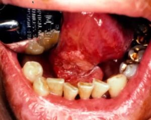
Here’s a little less obvious lesion. It’s kind of erosive like erythroplakia, regular, slightly raised leukoplakia but this is also a squamous cell carcinoma along the lateral border of the tongue.
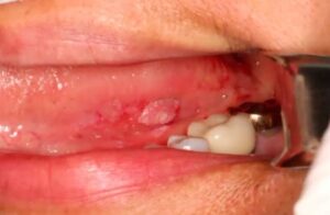
These are the things that you’re trying to look for in an earlier stage. You can recognize it when it’s just a red area or just a little white area. If you’re not sure if it was just a localized white area, smooth down these cusps and have the patient back in two weeks. If it’s not gone, you have to biopsy the lesion and make sure that it’s not dysplasia or a squamous cell.
Vesicular Ulcerative Conditions
Download the Diagnosing Vesicular Ulcerative Conditions checklist to get all the key information and images from this article.
Postgraduate Oral Pathology and Radiology Certificate
Learn more about the clinical and didactic skills necessary to evaluate and manage patients with oral diseases by enrolling in Herman Ostrow School of USC’s online, competency-based certificate program in Oral Pathology and Radiology.

