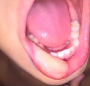
In this article we’ll review pictures from a patient that had a dental procedure and then a few days later noticed concerning ulcerations on their lip resulting from post-operative trauma.
Don’t have time to read this article? We get it. Download the Diagnosing Vesicular Ulcerative Conditions checklist to get the key information and images from this article plus all the other conditions we cover in the Dentist’s Guide to Oral Pathology.

It turns out that after the dental procedure, the mother took her child to get an ice pop. Because the child couldn’t feel their lip they left the ice pop there for so long that it necrosed the tissue.
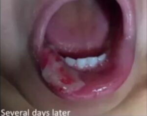
Then, several days later we see a traumatic ulceration secondary to epithelial necrosis. So you have necrosis, the tissue is dead, and it is noticeably different from the necrosis you might find in a patient suffering from Erythema Multiforme or the EM-TEN Spectrum.
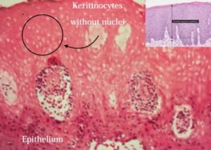
Interestingly you can see that the epithelium looks okay. You have your keratinocytes, but as you go towards the periphery you see that the keritinocytes have lost their nuclei which tells us that this epithelium is necrotic. This necrotic epithelium will eventually sloth and a traumatic ulcer in the mouth will form. That’s what we’re seeing here in this ulcerative area.
Bottom line here is make sure that you are informing the patient and their family that they need to wait to eat anything, even ice cream and ice pops, until they can feel their lip again. Post-op instructions are extremely important.
Traumatic Ulcerative Granuloma with Stromal Eosinophilia
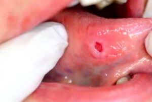
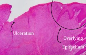
Here is another traumatic lesion of the oral cavity on the right lateral border of the tongue. We see it is a well-defined ulceration with slightly raised in white borders.
In the second image, we have a biopsy and see the overlying epithelium and the ulceration where there is no epithelium. It is covered by a fibrinopurulent membrane and there’s diffuse inflammation in the connective tissue.
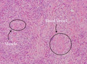
If you look closer you have a lot of these pink cells, which if you recall from our article on the Oral Pathology of Aphthous Ulcers and Crohn’s Disease, are eosinophils. There’s blood vessels, some muscle, and the ulceration is pretty deep. It’s going into the muscle. Zoom in and you can see eosinophils, neutrophils, and some fibroblasts in the ulcer bed that attempting to repair the granulation tissue.
How to Diagnose TUGSE
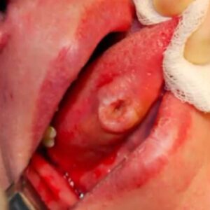
Here is a picture of a traumatic ulcer on the tounge.
This is something called a traumatic ulcerative granuloma. You can either have it secondary to trauma or it can be something we call TUGSE.
Traumatic Ulcerative Granuloma with Stromal Eosinophilia (TUGSE), means that there is a lot of eosinophils present. The main differential here is that it resembles squamous cell carcinomas. This lesion is concerning. One would think that this is a squamous cell, but you have to think about this entity as well.
For TUGSE, you would take a biopsy if it’s an ulcer of perilesional tissue. This happened to be a TUGSE. These are benign and reactive lesions, probably secondary to trauma.
How to Treat TUGSE
This will have been present for several months which makes us suspect squamous cell carcinoma. For treatment, once you biopsy the lesion it starts to resolve on its own with new blood flow in the area. It is chronic but it is self-limiting after the biopsy. Do not wait a couple weeks to take a biopsy.
Vesicular Ulcerative Conditions
Download the Diagnosing Vesicular Ulcerative Conditions checklist to get all the key information and images from this article.
Postgraduate Oral Pathology and Radiology Certificate
Learn more about the clinical and didactic skills necessary to evaluate and manage patients with oral diseases by enrolling in Herman Ostrow School of USC’s online, competency-based certificate program in Oral Pathology and Radiology.