This can be understood as the migration of virus from ganglion to surface along the course of sensory nerves. Upon reaching the surface the herpes virus infects epithelial cells and reproduces.
Secondary or recurrent herpes is something that most of us are familiar with. HSV is pretty ubiquitous. Only 12% of patients will have the symptomatic primary infection. Once the infection happens, the virus doesn’t get cleared it just lays dormant in your trigeminal ganglion.
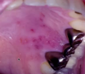
When the virus gets reactivated it goes from the ganglion, through the sensory nerves, and then to the surface. For recurrent herpes, you’re gonna see little vesicles so you can see them pretty well here.
Intraorally you may or may not see the vesicles because they might be subject to trauma. Intraorally you’re going to see recurrent herpetic lesions limited to attached mucosa. Recurrent herpes only sees it on the attached mucosa, and oftentimes it’ll be secondary. It will appear secondary to a cleaning in the area or some sort of trauma. There are patients who may have a prodromal syndrome or onset with paresthesia or pain or itching in the area 6 to 24 hours before the lesions actually develop.
Don’t have time to read this article? We get it. Download the Diagnosing Vesicular Ulcerative Conditions checklist to get the key information and images from this article plus all the other conditions we cover in the Dentist’s Guide to Oral Pathology.

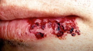
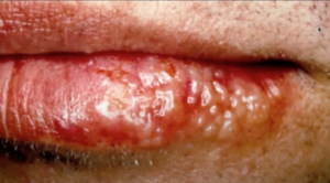
Here’s another picture of the early lesions of herpes labialis and a little bit later stage, a little bit more pristine.
Several days later they’re all becoming crusty. Remember, don’t touch these and then touch yourself or someone else because you can transmit the virus to other areas of your body.
Intraoral Recurrent Herpes
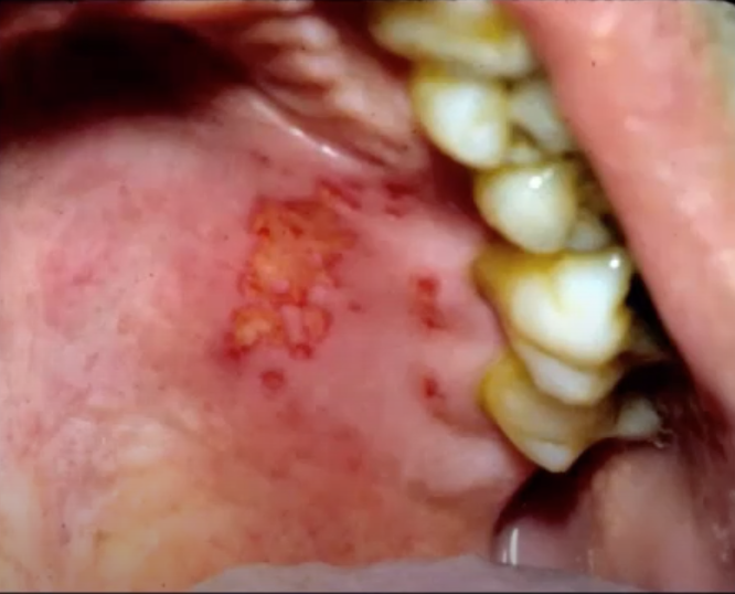
Here is another less vesicular, more ulcerative, example. that is a little bit further on in the process. If you biopsied this you would probably get a false negative. This is what herpes looks like in the oral cavity.
If you do take a biopsy of something like this on moveable mucosa and it comes back as herpetic this means this is an atypical presentation. What do you need to think about?
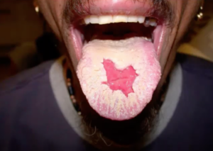
You need to think about the fact that the patient might be immune compromised. This is a patient that was undergoing chemotherapy for carcinoma and their immune system was compromised. Then they had recurrent herpes on moveable mucosa which is not seen in a normal, immunocompetent person.
Here is another example. This was taken by a friend of mine that lives in Texas and actually looks a lot like Texas so he was kind of excited about that. This patient had a known history of HIV and so this is again an atypical herpetic presentation.
Herpes in an immunocompetent individual is going to be limited to the keratinized mucosa or attach mucosa. If you see herpes and you can only really tell by biopsying moveable mucosa, then you’ve got to start working the patient up from an immunocompromised state.
Postgraduate Oral Pathology and Radiology Certificate
Learn more about the clinical and didactic skills necessary to evaluate and manage patients with oral diseases by enrolling in Herman Ostrow School of USC’s online, competency-based certificate program in Oral Pathology and Radiology.

