Oral precancerous lesions represent a significant public health challenge worldwide. These conditions, which include leukoplakia, erythroplakia, and oral submucous fibrosis, oral lichen planus among others, serve as potential precursors to oral cancer. With proper public health initiatives focused on early detection and intervention, many cases of oral cancer could be prevented.
Understanding Oral Precancerous Lesions
The World Health Organization (WHO) defines oral epithelial dysplasia (OED) as a “spectrum of architectural and cytological changes associated with an increased risk of progression to oral squamous cell carcinoma (OSCC)” [1]. Oral potentially malignant disorders (OPMDs) encompass clinical presentations that may precede OSCC diagnosis.
Recent literature indicates that OED (Figure 1) and squamous cell carcinoma are present in 40–46% of leukoplakia cases [2,3]. This high percentage underscores the importance of thorough evaluation and monitoring of oral white lesions.
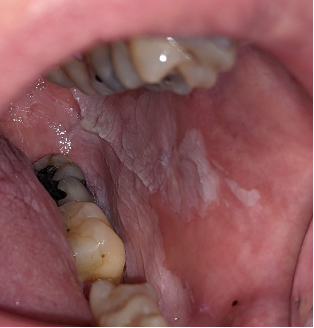
Fig 1: Oral epithelial dysplasia (OED) on the left buccal mucosa
Like what you’re learning? Download a brochure for our online, postgraduate Oral Pathology and Radiology certificate program.
Global Burden and Risk Factors
The global prevalence of oral leukoplakia is approximately 4.11% [3,4]. Malignant transformation rates vary according to the severity of dysplasia:
- Mild epithelial dysplasia: 11.9-18.1%
- Moderate epithelial dysplasia: 8.7-17.9%
- Severe epithelial dysplasia: 32.2-60% [5]
Risk factors include tobacco use, alcohol consumption, HPV infections, and poor oral hygiene. South Asia bears a disproportionate burden due to widespread use of betel quid and tobacco products, while in Western countries, HPV infections and alcohol consumption are primary contributors [6].
Public Health Initiatives
Screening Programs: Community-based screening initiatives have proven effective in identifying oral precancerous lesions early. These programs often utilize:
- Mobile screening units with imaging technology
- Visual and fluorescence-assisted examinations
- Free or subsidized check-ups for high-risk populations [7]
Research shows that countries with robust screening programs report increased early detection rates, leading to improved survival rates [8].
Educational Campaigns: Public awareness forms the cornerstone of prevention strategies. Effective educational approaches include:
- Government and NGO-led awareness campaigns
- Social media and traditional media outreach
- Integration of oral health education in school curricula [9]
Tobacco and Alcohol Control: Countries implementing strict tobacco and alcohol regulations have observed declining rates of oral precancerous lesions. Effective measures include:
- Higher taxation on tobacco and alcohol products
- Graphic health warnings on packaging
- Smoke-free legislation [10]
Technological Advances in Detection and Treatment
Diagnostic Innovations: Emerging technologies are transforming the detection landscape:
- AI-assisted imaging tools for analyzing lesions
- Portable fluorescence-based devices for non-invasive detection
- Digital pathology enabling remote analysis of biopsy samples [11]
Institutions like USC are opening new facilities for oral pre-cancer lesions and working in collaborative research between the Ostrow School of Dentistry and the Keck School of Medicine to predict the malignant transformation of oral leukoplakia, potentially revolutionizing risk assessment and treatment planning.
Treatment Approaches: Current treatment options include:
- Surgical excision or laser ablation
- Watch-and-wait approach for lower-risk lesions
- Immunotherapy (emerging option)
- Off-label use of medications like imiquimod 5% [12]
Recent clinical trials investigating nivolumab for high-risk oral leukoplakia have shown potential for immunotherapy as a non-invasive treatment option [13]. While still experimental, these approaches offer hope for patients with extensive or multifocal disease.
Challenges and Future Directions
Despite progress, significant challenges remain:
- Limited funding for public health initiatives in developing countries
- Healthcare workforce shortages in rural areas
- Cultural stigmas preventing individuals from seeking care
- Lack of standardized management guidelines [14,15]
Future efforts should focus on:
- Developing evidence-based treatment protocols
- Increasing funding for prevention programs
- Training more healthcare providers in oral pathology
- Conducting clinical trials with histopathological assessment
- Addressing healthcare disparities in vulnerable populations
Conclusion
Oral precancerous lesions present a preventable global health challenge. Through coordinated public health efforts, including early detection programs, awareness campaigns, and innovative treatment approaches, the burden of oral cancer can be significantly reduced. A multidisciplinary approach involving governments, healthcare providers, researchers, and international organizations is essential to ensuring equitable access to preventive care and treatment worldwide.
Postgraduate Oral Pathology and Radiology Certificate
Learn more about the clinical and didactic skills necessary to evaluate and manage patients with oral diseases by enrolling in Herman Ostrow School of USC’s online, competency-based certificate program in Oral Pathology and Radiology.
References
- World Health Organization. (2017). WHO classification of head and neck tumours (4th ed.). IARC Press.
- Warnakulasuriya S, et al. (2016). “Oral epithelial dysplasia classification systems: predictive value, utility, weaknesses and scope for improvement.” Journal of Oral Pathology & Medicine, 45(6), 367-373.
- Petti S. (2003). “Pooled estimate of world leukoplakia prevalence: a systematic review.” Oral Oncology, 39(8), 770-780.
- Müller, S. (2018). Oral epithelial dysplasia, atypical verrucous lesions and oral potentially malignant disorders: focus on histopathology. Oral Surgery, Oral Medicine, Oral Pathology and Oral Radiology, 125, 591–602.
- Chaturvedi, A.K., et al. (2020). Oral Leukoplakia and Risk of Progression to Oral Cancer: A Population-Based Cohort Study. Journal of the National Cancer Institute, 112(10), 1047-1054.
- Speight, P.M. (2007). Update on oral epithelial dysplasia and progression to cancer. Head and Neck Pathology, 1(1), 61-66.
- Warnakulasuriya, S. (2009). Global epidemiology of oral and oropharyngeal cancer. Oral Oncology, 45(4-5), 309-316.
- Sankaranarayanan, R., et al. (2013). Cancer screening in India: strategies and outcomes. Journal of Cancer Research and Therapeutics, 9, 402-406.
- Brocklehurst, P., et al. (2013). Screening programmes for the early detection and prevention of oral cancer. Cochrane Database of Systematic Reviews, (11).
- Petersen, P.E. (2009). Oral cancer prevention and control – The approach of the World Health Organization. Oral Oncology, 45(4-5), 454-460.
- Gallus, S., et al. (2013). European Code against Cancer 4th Edition: Tobacco and cancer. Cancer Epidemiology, 39, S20-S33.
- Vistoso Monreal, A., et al. (2023). Artificial intelligence for the prediction of malignant transformation of oral leukoplakia. Oral Surgery, Oral Medicine, Oral Pathology and Oral Radiology. https://doi.org/10.1016/j.oooo.2023.07.030
- Martinez-Lopez, A., et al. (2017). Successful treatment of proliferative verrucous leukoplakia with 5% topical imiquimod. Dermatologic Therapy, 30:e12413.
- Hanna, G.J., Villa, A., et al. (2024). Nivolumab for Patients With High-Risk Oral Leukoplakia: A Nonrandomized Controlled Trial. JAMA Oncology, 10(1), 32-41.
- Awadallah, M., et al. (2018). Management update of potentially premalignant oral epithelial lesions. Oral Surgery, Oral Medicine, Oral Pathology and Oral Radiology, 125(6), 628-636.