A biopsy is defined as the sampling or removal of tissues or liquids from the body for examination, in order to determine the existence or cause of a disease. A biopsy is strongly recommended for most of the lesions that persist for more than two weeks which interferes with oral function, or does not improve by removing the local irritants.
Like what you’re learning? Download a brochure for our online, postgraduate Orofacial Pain and Oral Medicine degree program.
6 Basic Etiologic Categories for an Oral Lesion
Whenever you see an oral lesion, train yourself by using the diagnostic sieve to list out possible clinical differential diagnoses:
- Developmental (Congenital)
- Inflammatory/Infection
- Neoplastic
- Traumatic
- Autoimmune/Allergic
- Oral manifestation of systemic disease
Related Reading: The Dentist’s Guide to Oral Pathology of Vesicular Ulcerative Conditions
Don’t have time to read The Dentist’s Guide to Oral Pathology of Vesicular Ulcerative Conditions? Download the checklist!
6 Reasons Why It Is Important to Have a Differential Diagnosis
- It helps you determine when to perform the biopsy: Is it urgent?
- It helps you determine how to perform the biopsy: Punch, Incisional or Excisional biopsy? What instruments do I need?
- It is how doctors approach diagnosis of an unknown disease.
- It will demonstrate to the patient, third party, and pathologist that you have included all the clinical possibilities.
- It generally leads to better decision-making.
- It also helps to prevent errors in judgment and misdiagnosis or failure to diagnose.
What kinds of biopsies are available?
1. Punch Biopsy
Punch biopsies are commonly used in dermatology for sampling of skin lesions. It is also used for gingival biopsies especially for cases of pemphigus vulagaris or mucous membrane pemphigoid.
2. Excisional Biopsy
An excisional biopsy removes the entire lesion and it is both a therapeutic as well as a diagnostic procedure. Excisional biopsies are most commonly used for lesions of 1 cm or less, for even larger benign lesions to avoid multiple surgeries, or when complete removal is possible without significant morbidity.
3. Incisional Biopsy
An incisional biopsy involves taking a small portion of the lesional tissue for diagnostic purpose. Incisional biopsies are commonly used:
- When a lesion is large enough that definitive removal for histologic diagnosis would produce significant morbidity.
- When necessary to convince a patient that serious pathology exists although the patient may not agree or may be asymptomatic.
- When malignancy is highly suspected and biopsy is performed for confirmation and rapid diagnosis rather than attempting to remove the cancer completely with clear margins.
Additional Reading: Infectious Lesions of the Oral Cavity: Histoplasmosis & Mucormycosis
What are the contraindications for an oral biopsy?
There are no contraindications for a biopsy when the risk of doing nothing outweighs the risk of the surgical procedure. In some instances, contraindications to surgical biopsy may exist due to underlying systemic conditions such as severe and uncontrolled hypertension. In such situations, the procedure can be delayed until appropriate precautions are in place such as:
- Asking the physician to lower the patient’s elevated blood pressure with medications.
- Asking the physician to give the patient clotting factor or stop anticoagulant medications if the patient has an elevated INR.
- Treating the local infection with antibiotics.
- Referring to the adequate institute to perform the biopsy in a high risk cardiovascular disease patient (e.g. hospital).
If you are confident the lesion is a malignancy, refer to the surgeon that is likely to manage, such as head and neck surgeon or oncologist.
Note: do not try to excise a malignancy during a biopsy as there will be no lesion left for the surgeon to assess for further treatment.
Like what you’re learning? Download a brochure for our online, postgraduate Orofacial Pain and Oral Medicine program.
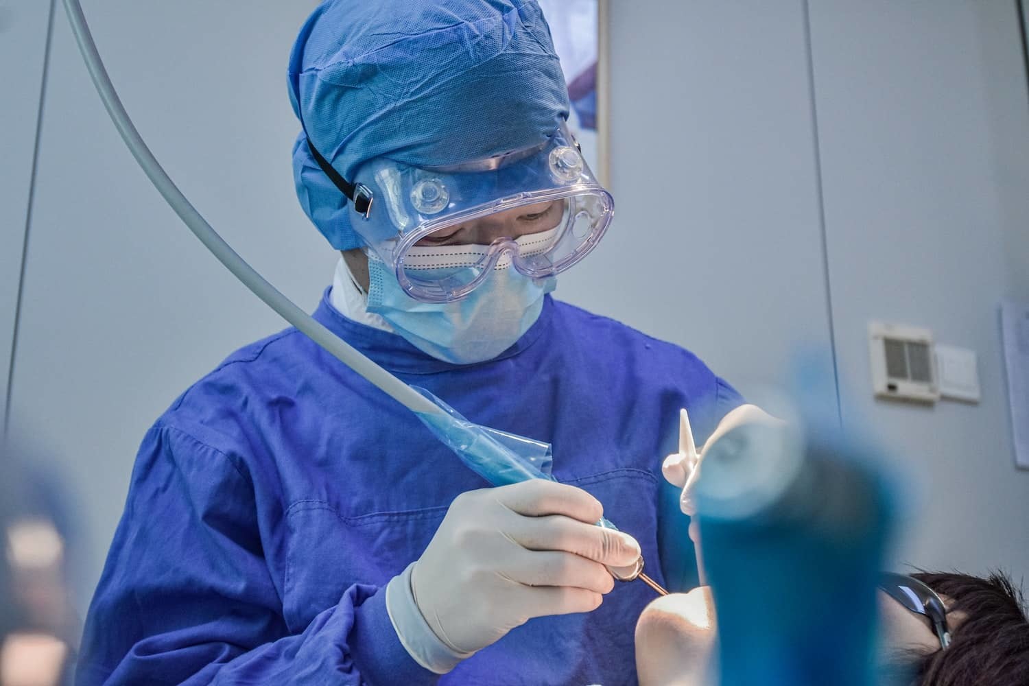
Step-by-Step Oral Biopsy Procedure
1. Select the Area to Biopsy
Prior to the procedure, you must have an idea of how, where, and what kind of procedure you are going to perform.
When pemphigus vulgaris or mucous membrane pemphigoid are suspected, the biopsy should be performed at the edge of the lesion to include both lesional and normal tissues because there may not be much normal tissue left at the center of the lesion due to a very thin epithelium or ulcer.
If the lesion is a precancerous lesion like leukoplakia or an early Squamous Cell Carcinoma (SCC) then you can biopsy just the center of the lesion and leave the border alone so that the lesion can be excised in total at a future appointment. You can also elect to biopsy a small part of the margin of the lesion and the normal tissue so that the pathologist can compare the normal and abnormal tissue.
Note: Early SCC may look like an erythroplakia or leukoplakia.
Related Reading: Oral Pathology of Oropharyngeal Squamous Cell Carcinoma
2. Prepare the Instruments and Environment
The basic instruments you need for a soft tissue biopsy include:
- Local anesthetic cartridge and syringe
- Scalpel: Blade handle with the blade or a disposable scalpel (No. 15 and 12 blades are most commonly used)
- Tissue forceps with and without teeth
- Retractors
- Needle holder and suture (4-0 or 5-0 silk sutures are commonly used intraorally)
- Scissors
- Gauze
- Curved forceps, Bite block (as needed)
- Specimen bottle with fixing solution and biopsy data sheet
3. Administer Anesthesia
Use both topical and local anesthesia in an attempt to obtain less or no pain to the patient during the procedure. In general, patients are afraid and nervous for any surgical procedures. The more pain control you achieve, the greater the chance of success. For large lesions, anesthesia blocks are preferable. Also, if you are planning to perform an incisional biopsy, do not infiltrate inside the lesion as that may create artifacts (vacuolation).
Do Not Rush to the Next Step
Take enough time for the local anesthesia to work. You will not only achieve good pain control, but excellent hemostasis as well, which will ultimately make the procedure easier.
4. Make a Wedge-Shaped Incision
For both incisional and excisional biopsies, a wedge-shaped incision is desired in an attempt to have a clean, desirable closure. The incision should extend beyond the suspected depth of the lesion.
Biopsy Incision Tips
- If possible, include the adjacent normal tissue.
- Do not use small strokes with the scalpel. Incisions should be continuous. Small strokes will destroy the cell alignment of the tissue and the orientation.
- Do not cut the specimen to see what might be inside. The pathologist may want to ink the specimen to establish the margins.
- For excisional biopsies, take enough representative tissue without damaging the tissue integrity.
- Be aware of adjacent anatomy to avoid unnecessary tissue damage.
- Be cautious not to crush the specimen with tweezers or suction the specimen. Accidental suctioning of tissue is very common. (Educate the assistant well prior to the procedure).
Note: Length of incisions should be parallel to natural resting lines when possible to optimize esthetics.
Like what you’re learning? Download a brochure for our online, postgraduate Orofacial Pain and Oral Medicine program.
5. Remove and Transfer the Specimen
A. Use One Bottle Per Specimen
Do not mix specimen unless it is a connective tissue mass that came out in several parts.
B. Fill with Formalin
The volume of formalin should be at least 20 times the volume of the specimen. If immunofluorescence analysis is desired, the specimen needs to be placed in a different solution called Michel’s solution.
C. Label the Bottle
Each container should be identified with the patient’s name, clinician’s name, date and the site of the biopsy.
D. Stabilize the Tissue
Place mucosal or epithelial biopsies on a small rigid piece of paper before placing in the formalin bottle to prevent the tissue from rolling around itself and collapsing which would make it more difficult for the pathologist to orient and interpret the specimen.
E. Place an Orientation Suture (if required)
When performing a biopsy of a suspicious lesion (possible tumor), place an orientation suture in the specimen (one suture will be enough) so the pathologist can tell you which margins are clear or not.
F. Transfer to the Transport Media
Put the sample immediately into the transport media, not on gauze or on the surgical tray where it may dry out or undergo significant autolysis, and make sure the cap is tight so it does not spill while being transported to the pathology lab.
G. Fill out the Biopsy Data Sheet
Provide the pathologist with clinical and background information including: clinical appearance, duration, symptoms, pertinent medical history, risk factors, and clinical differential diagnosis.
If possible, submit copies of clinical or radiographic images with the biopsy to give the pathologist the ability to correlate clinical, radiographic, and histopathologic findings.
Lastly, do not draw on or circle the lesion on photographs or radiographs. The pathologist will be able to see the abnormality without you pointing it out and possibly distorting the image.
6. Obtain Hemostasis and Wound Closure
Best hemostasis can be obtained by direct pressure unless you damaged a larger vessel.
For patients who will definitely need follow-up, use non-resorbable silk sutures. One suture should be placed every 3-5mm’s, and the wound should be sutured without tension to avoid dehiscence. Similarly, do not pull the knot too tight for soft tissue suturing.
Note: Do not try to achieve hemostasis by using anesthesia.
7. Provide Post-operative Instructions
Review the aftercare instructions with the patient and preferably a family member to ensure adherence and understanding.
A. Pain
If the procedure site was large or if the patient is already experiencing pain, prescribe NSAIDs or stronger analgesics (e.g. Vicodin). For small procedures, patient can take OTC analgesics such as a 200-mg tablet of Ibuprofen (Advil, Motrin) 3 times a day with food, or a 325-mg tablet of Acetaminophen (Tylenol) every 6 hours, or as needed.
For any biopsy, instruct the patient to take one tablet of analgesic before the anesthesia wears-off. If you do not hold any analgesics in your office, you should advise the patient to bring in the medication which they usually take when they have pain at the day of biopsy.
B. Bleeding
Bleeding might occur especially during the first few days: Patient can apply direct pressure for 10 minutes using a gauze or tissue to stop bleeding. If it continues to bleed, instruct patient to contact the clinic.
C. Swelling and Bruising
Swelling can happen as a healing process. Educate the patient that peak swelling is generally the 3rd or 4th day after the procedure and then will gradually subside. Bruising is less common for soft tissue biopsies, but might happen depending on the extent of the procedure and the patient’s age (e.g. older) or immunity.
D. Smokers
For smokers, educate them to stop smoking during the time when tissue is healing (at least a week).
E. Additional Biopsies
Lastly, inform the patient the possibility of another biopsy in the future depending on the diagnosis the pathologist makes. If the histologic diagnosis does not seem correct with your clinical impression, talk to the pathologist personally and decide if you need another biopsy or not.
Further Reading
- Oral Pathology of Oral Pharyngeal Squamous Cell Carcinoma & HPV
- Aphthous Stomatitis: Treatment, Diagnosis, and Clinical Pictures
- Oral Pathology of Aphthous Stomatitis and Crohn’s Disease
- What is Erythema Multiforme and the EM-TEN Spectrum?
- Understanding Oral Herpes: Primary Herpetic Gingivostomatitis
- Oral Pathology of Secondary (Recurrent) Herpetic Eruptions
- Oral Pathology of Primary, Secondary, and Tertiary Syphilis
Earn an Online Postgraduate Degree in Orofacial Pain and Oral Medicine
Like what you’re learning? Consider enrolling in the Herman Ostrow School of Dentistry of USC’s online, competency-based certificate or master’s program in Orofacial Pain and Oral Medicine.