We are going to review the differences between vesicles and ulcerations, two types of lesions found in an oral cavity. If you look at this oral mucosa decision tree for oral lesions, many of these lesions are classified as vesiculobullous or vesicular ulcerative conditions.
Don’t have time to read this article? We get it. Download the Diagnosing Vesicular Ulcerative Conditions checklist to get all the key information and images from this article plus all the other conditions we cover in the Dentist’s Guide to Oral Pathology.

The Difference between a Vesicle and an Ulcer
In order to explore the scope of ulcerative conditions in later articles, let’s start with the difference between a vesicle and an ulcer.
What is a vesicle?
A vesicle is an elevated, fluid-filled lesion covered by epithelium. The fluid may accumulate either within the epithelium (intraepithelial) or below the epithelium (subepithelial). It is a small bulla, less than or equal to 1 centimeter in diameter.
What is an ulcer?
An ulcer is a defect or disruption in the continuity of the epithelial component of the oral mucosa so that a depression exists. This is often covered by a fibrinopurulent membrane. A vesicle may eventually lose the overlying epithelium and then present as an ulceration.
Differential Diagnosis of a Vesicle and Oral Ulcer
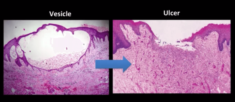
The left photomicrograph shows fluid accumulation within the stratified squamous epithelium, which causes an ovoid defect. Also, note the presence of inflammation in the underlying connective tissue.
As you can imagine, the superior aspect of the vesicle, the thin compromised portion of the epithelium is very fragile and friable. When this is subject to trauma it disintegrates and the overlying epithelium is lost. The result is an ulceration.
The right photomicrograph shows the absence of stratified squamous epithelium centrally. At either edge is hyperplastic and hyperparakeratotic stratified squamous epithelium. The ulcer bed is composed of granulation tissue, inflammatory cells, and a lot of congested blood vessels.
Now that we understand the difference between a vesicle and an ulcer, we’ll look at more complex ulcerative conditions. In the next article, we’ll cover diagnosing, treating, and reviewing histological differentials of pemphigus vulgaris.
Don’t forget to download the Diagnosing Vesicular Ulcerative Conditions checklist for all the key information from this article and the Dentist’s Guide to Oral Pathology.