
Four things to measure in a patient with centralized orofacial pain, and why the pain score alone will mislead you.
A patient with chronic masticatory pain starts nortriptyline. Six weeks later you ask how the pain is. She says it is about the same. Do you add gabapentin, or has the drug not been given a fair trial?
This is the commonest decision in our clinic, and we usually make it on a single number. Unfortunately, that number is the weakest instrument we own. Global pain ratings (0–10) are noisy, they carry the whole week’s mood and sleep with them, and they are the last thing to move. A muscle palpation score may change before the patient’s summary of the month does.
This piece is about what instruments a clinician can use and write down. It is not a research protocol, and it is not quantitative sensory testing in the laboratory sense. It is four measurements you can take in about five minutes at every visit, using equipment you already have.
Why the pain score is the wrong single instrument
Once a peripheral pain has persisted long enough, it can begin to sensitize the pathway that carries the signal to the cortex. This is called centralized pain, but it is not one thing going wrong, it is two, and the two are separate. First, the system can amplify what arrives from the periphery. Amplification is what temporal summation measures: repeat an identical stimulus and, if it is being amplified, it hurts progressively more. Second, the system can fail to dampen the signal as it ascends. Failed damping is what conditioned pain modulation (CPM) measures: pain applied somewhere else should reduce pain here, and does not. A given patient can be abnormal on either, on both, or on neither.
Nortriptyline and duloxetine act on the descending inhibitory synapse. Gabapentin acts presynaptically on the calcium channel and reduces excitatory transmitter release. Two mechanisms, two targets. There is no validated chairside test that tells you which one is broken in the patient in front of you, which is why our protocol steps these medications in sequence rather than trying to identify the broken mechanism and then select the logical drug. A tricyclic first, with gabapentin added in non-responders, is also the only published sequence in this population (Haviv et al. 2015).
But stepping only works if you can tell what a response looks like. And a global pain score (0–10) has three problems as that signal. It aggregates everything. It is anchored to the patient’s memory of an average week. And with a slow drug it is the last measure to move.
| The finding that should change your chart note In the only placebo-controlled trial of gabapentin in chronic masticatory myalgia (Kimos et al. 2007; 50 randomized, 36 completed, 12 weeks), the muscle measure moved a month before the pain score. The tender site count separated from placebo at week 8 and stayed separated at week 12 (P < .001). Patient-reported pain did not separate until week 12 (P = .026). Both arms began at 9.50 tender sites out of 12. By week 12 the gabapentin arm was at 3.04 and placebo at 7.60 — a reduction of 6.46 against 1.90 (P = .002). In the repeated-measures analysis the time-by-group interaction reached significance only for the tender site count (P = .004), not for either pain scale. It was the only measure whose trajectories genuinely diverged. Note what was in the examiner’s hand. Those counts were made with an algometer scored against normative thresholds for each site, not with fingers. |
Week 8 is roughly where you are deciding whether to add a second drug. Below is what to do with that.
The five-minute panel
Take these at baseline, at four to six weeks, and again when the patient reaches a stable dose. Same examiner, same sites, same order, roughly the same time of day.
| Measure | How to do it | What it tells you, and what it does not |
| Tender site count | Conduct a standard muscle palpation. Record the number of sites that are painful, not just whether the patient has myalgia. | The best supported of the four, and the only one shown to move before the pain score does: in the one placebo-controlled gabapentin trial in masticatory myalgia it separated at week 8, the pain score at week 12 (Kimos et al. 2007). Costs nothing, and you are already collecting it. |
| Spread on palpation | Assess how far the pain spreads under sustained pressure rather than judging whether it is referred. Hold the most tender point for 5 seconds. Have the patient shade where they feel it on a printed face-and-neck diagram. Count the areas shaded. | The measure that speaks most directly to central amplification. It is also the least validated of the four in the face: spread beyond the palpated site is common in healthy people under standardized palpation (Masuda et al. 2018). Treat a shrinking map as encouraging, not as proof. |
| Pressure pain threshold | An algometer is best: a 1 cm² probe tip, pressure applied at a constant rate until the patient reports pain. Two readings per site, masseter and anterior temporalis, both sides. | Highly repeatable, so a real change is interpretable. But it moves with peripheral tissue state as well as central state, so it is not a central marker on its own. |
| Temporal summation | Weighted pinprick. Rate one stimulus, then rate the peak of ten delivered at one per second. Record the difference between the two ratings. | The closest thing to a bedside read on facilitation. Record the difference, not the ratio: in meta-analysis the absolute change was substantially more reliable than the ratio (Chen et al. 2025). |
1. Count the tender sites
You are already palpating. The change is to record a count rather than a category. A patient who went from eleven tender sites to five has improved in a way that is legible to you and to the next clinician, even if she still says her pain is a six. This is the measure with the strongest supporting data in our own field, and the only one shown to declare itself before the pain score does (Kimos et al. 2007). It costs nothing but a little time.
One practical footnote. In that trial the count was made with a rate-controlled algometer, and a site scored positive only when the reading fell below a published normative threshold for that site. If your clinic owns an algometer, use it for the count as well as for the threshold — that is how the result was produced, and it removes you as a variable. Fingers will still show you the direction; they will not reproduce the trial.
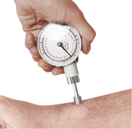
Figure 1. Tender site count. A standard pressure is applied to each site — superficial and deep masseter, anterior temporalis, and thenar — and the number of painful sites is recorded. A rate-controlled algometer removes the examiner as a variable.
Like what you’re learning? Download a brochure for our Orofacial Pain and Oral Medicine certificate or master’s degree program.
2. Map the spreading pain
A note on the word. We have removed “referred pain” from our clinical lexicon and use “spreading pain” instead. Deciding whether a pain is truly referred is close to impossible to define reproducibly; in practice it is done by a kind of intuition that does not survive a second examiner. Where the literature cited below says referred pain, read it as spread. What is reproducible is not whether the pain is referred, but how far it goes.
Healthy people report spread readily when you press hard enough: standardized palpation of the masseter at 0.5 to 2.0 kg evoked spreading pain or sensations in pain-free volunteers (Masuda et al. 2018). So do not record spread as present or absent. Record how far it goes, using a steady pressure of about 1 kg held for 5 seconds, and keep it as a picture.
Area is the variable that separates patients from controls. When spread was drawn on anatomical maps, digitized and measured in 25 women with myofascial temporomandibular disorder against 25 matched healthy women, with the examiner blinded to which was which, the areas were significantly larger in the patients (Fernández-de-las-Peñas et al. 2010). That is the best published argument for grading how far the pain goes rather than whether it goes anywhere at all.
Hold the most tender point with sustained pressure for five seconds and have the patient shade, on a printed face-and-neck diagram, everywhere she feels it. Keep the diagrams in the chart. Over three visits you get a series of maps, and a shrinking map is the most direct clinical sign you have that central amplification is settling.
Two honest limits. First, the pressure your finger applies is not standardized, so some of what you see between visits is your own variation rather than the patient’s nervous system. Using an algometer to deliver the sustained stimulus removes most of that — and note that the study above used manual palpation, so it shares the limitation. Second, spread is not a pure central sign: a muscle with an active local generator spreads too. Patients whose pain spreads beyond the muscle border do have substantially more widespread pain than those whose pain stays local, but the authors of that work conclude the sign is not yet established as a valid clinical test for central sensitization (Lövgren et al. 2022). Read a shrinking map as encouraging, not as proof.
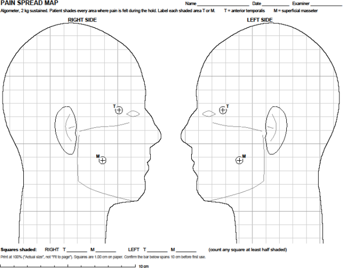
Figure 2. Pain spread map. The patient shades every area where pain is felt during the five-second sustained load. Shaded squares are counted and the sheet is filed in the chart, so the record is the picture rather than a description of it.
3. Pressure pain threshold, at the same marked sites
Two readings each at masseter and anterior temporalis, both sides, with a constant-rate algometer. This is the most repeatable measure in the panel, which is what makes a change in it worth believing. It also has the clearest interpretation problem: threshold rises when the muscle calms down, whatever calmed it. It is a good measure of whether the patient is better and a poor measure of why.
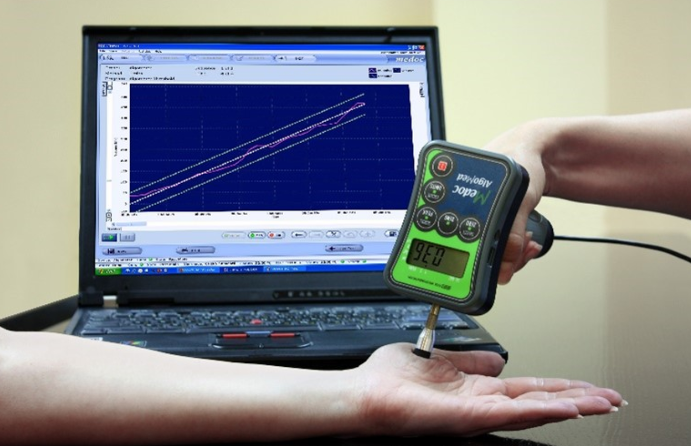
Figure 3. Pressure pain threshold. A 1 cm² probe tip with pressure rising at a constant rate until the patient first calls it painful. Two readings at each marked site, every visit.
4. Temporal summation with a weighted pinprick
Rate a single pinprick, then rate the peak of ten delivered at about one per second, and record the difference between the two ratings. This is the one item in the panel that speaks directly to facilitation rather than to sensitivity in general, and it takes under two minutes.
Two practical notes, both from the same recent meta-analysis of temporal summation reliability (Chen et al. 2025). First, record the absolute difference — the rating on the tenth stimulus minus the rating on the first — and not the wind-up ratio: pooled reliability was good for the absolute change (ICC 0.81) and poor for the ratio (ICC 0.36). Small-contact-area mechanical stimulation at 1 Hz was the most dependable form. Second, and this is the limit to know before you act on the number: between-session reliability of temporal summation was poor overall (ICC 0.49), even in healthy volunteers. Repeating it across visits is exactly the use proposed here, so treat a change in this item as the weakest of the four and do not let it outvote the palpation findings.
Figure 4. Temporal summation. A weighted pinprick, rated once, then delivered ten times at about one per second with the peak rated again. Record the difference between the two ratings, not the ratio.
What about von Frey hairs and allodynia?
We routinely look for allodynia, but it is difficult to measure. In the hand or foot with cutaneous neuropathic pain you can quantify it with a von Frey filament, but muscle pain is not cutaneous pain, and using von Frey filaments intraorally is problematic. For now we do not recommend von Frey measurements. Use pressure pain threshold for persistent muscle pain, and the temporal summation method described above.
Dynamic mechanical allodynia, stroking with a brush, is quick and clinically vivid. Be careful about relying on the patient’s report of it. In 617 patients with neuropathic pain, self-reported dynamic allodynia agreed with bedside examination at kappa 0.39 and with laboratory sensory testing at kappa 0.17, with an area under the curve of 0.65 (Gierthmühlen et al. 2019). Elicit it yourself, or do not record it.
What this panel is, and what it is not
None of these four tests is quantitative sensory testing as a laboratory would define it, and the panel is not offered as a substitute. Formal testing of descending inhibition requires a conditioning stimulus, a cold pressor or a pressure cuff, and roughly twenty minutes, and it has a reliability problem of its own that is not yet solved.
A simplified bedside kit is not a fantasy. An algometer, a pinprick and a conditioning clamp have been assembled into a three-item bedside battery with acceptable test-retest reliability and assessment validity against laboratory testing in knee osteoarthritis (Izumi et al. 2022). No orofacial equivalent has been built. In a dental office, an algometer and a weighted pinprick are the practical core.
| Read this before you use the panel to change a prescription The panel has not been validated as a treatment-response measure in orofacial pain. The tender-site evidence rests on one study, and that study enrolled 50 women aged 18 to 45 and reached a mean gabapentin dose above 3,300 mg a day — roughly three times what most of us prescribe. Its direction transfers to your patient; its numbers do not. The pressure threshold and temporal summation evidence comes largely from other body regions. The spread map has no normative data for the face and no published reliability figures for facial muscles. It is offered as a structured way to observe your own patient over time, not as a test with a cutoff. A measurement that improves while the pain score does not is a reason to give the drug more time. It is not proof that the central nervous system has changed. |
Reading the panel against the clock
The reason to collect this at all is that our drugs are slow and patients’ pain memory is poor. That makes an early pain score helpful but unreliable.
Nortriptyline. Reuptake blockade is immediate. What takes weeks is receptor adaptation and the rise in tonic noradrenergic output. Allow four to six weeks at an adequate dose before calling it a poor response. No trial has established that interval for a tricyclic in masticatory myalgia; it is a reasoned schedule, not a measured one. The nearest thing to a measured one is the gabapentin trial above, where the muscle measure declared itself at week 8 and the pain score not until week 12.
Gabapentin. It is not a channel blocker. Chronic, not acute, exposure reduces the number of α2δ calcium channel subunits reaching the nerve terminal, so the effect appears only with sustained dosing and the subunits already installed have to turn over (Hendrich et al. 2008). Allow two weeks of titration, then four to six weeks at dose. That mechanism comes from expression systems and dorsal root ganglion neurons rather than from patients, and it is not universally replicated.
End to end, the stepped sequence takes ten to fourteen weeks. Most drugs called failures in this clinic were stopped at four.
A week-one report that it is already helping is usually sleep rather than analgesia. Do not judge either agent on it, in either direction.
Against that timetable, four numbers measured over time are worth considerably more than one number taken once. If the tender site count is falling and the spread map is shrinking at week six while the pain score has not budged, the drug is doing something and the patient should stay on it. If nothing has moved on any of the four, adding a second agent is the right call — and now you have a reason on paper.
A note on the numbers we quote
Two things are worth saying plainly, because they get repeated as though they were settled. First, the timeline. We tell patients three to six months for channels and receptors to turn over, assuming they take the medication consistently. That figure is a reasoned extrapolation from animal and cell work, not a measured schedule in sensitized human nerve. It is also not a protein half-life: the turnover half-life of neuronal sodium channels is reported to be on the order of one to three days (Devor 2006). The months are the expression program reverting, not the individual molecules aging out.
Second, drug efficacy. The current meta-analysis of neuropathic pain pharmacotherapy puts the number needed to treat at 4.6 (95% CI 3.2–7.7) for tricyclics, 7.4 (5.6–10.9) for serotonin-noradrenaline reuptake inhibitors, and 8.9 (7.4–11.1) for gabapentinoids (Soliman et al. 2025). Those are neuropathic pain figures, not temporomandibular disorder figures, and they are more sobering than the numbers many of us learned. They are also the reason to measure carefully rather than to keep switching drugs.
Are you interested in a variety of issues focused on orofacial pain, medicine and sleep disorders? Consider enrolling in the Herman Ostrow School of Dentistry of USC’s online, competency-based certificate or master’s program in Orofacial Pain and Oral Medicine.
References
Chen KK, Rolan P, Hutchinson MR, de Zoete RMJ. Reliability of temporal summation of pain in healthy and clinical populations: a systematic review and meta-analysis. Eur J Pain 2025;29(8):e70097.
Devor M. Sodium channels and mechanisms of neuropathic pain. J Pain 2006;7(1 Suppl 1):S3–S12.
Fernández-de-las-Peñas C, Galán-del-Río F, Alonso-Blanco C, Jiménez-García R, Arendt-Nielsen L, Svensson P. Referred pain from muscle trigger points in the masticatory and neck-shoulder musculature in women with temporomandibular disorders. J Pain 2010;11(12):1295–1304.
Gierthmühlen J, Schneider U, Seemann M, et al. Can self-reported pain characteristics and bedside test be used for the assessment of pain mechanisms? An analysis of results of neuropathic pain questionnaires and quantitative sensory testing. Pain 2019;160(9):2093–2104.
Haviv Y, Rettman A, Aframian D, Sharav Y, Benoliel R. Myofascial pain: an open study on the pharmacotherapeutic response to stepped treatment with tricyclic antidepressants and gabapentin. J Oral Facial Pain Headache 2015;29(2):144–151.
Hendrich J, Van Minh AT, Heblich F, et al. Pharmacological disruption of calcium channel trafficking by the α2δ ligand gabapentin. Proc Natl Acad Sci U S A 2008;105(9):3628–3633.
Izumi M, Hayashi Y, Saito R, Oda S, Petersen KK, Arendt-Nielsen L, Ikeuchi M. Detection of altered pain facilitatory and inhibitory mechanisms in patients with knee osteoarthritis by using a simple bedside tool kit (QuantiPain). Pain Rep 2022;7(3):e998.
Kimos P, Biggs C, Mah J, Heo G, Rashiq S, Thie NMR, Major PW. Analgesic action of gabapentin on chronic pain in the masticatory muscles: a randomized controlled trial. Pain 2007;127(1–2):151–160.
Lövgren A, Visscher CM, Lobbezoo F, Yekkalam N, Vallin S, Wänman A, Häggman-Henrikson B. The association between myofascial orofacial pain with and without referral and widespread pain. Acta Odontol Scand 2022;80(7):481–486.
Masuda M, Iida T, Exposto FG, Baad-Hansen L, Kawara M, Komiyama O, Svensson P. Referred pain and sensations evoked by standardized palpation of the masseter muscle in healthy participants. J Oral Facial Pain Headache 2018;32(2):159–166.
Soliman N, Moisset X, Ferraro MC, et al; NeuPSIG Review Update Study Group. Pharmacotherapy and non-invasive neuromodulation for neuropathic pain: a systematic review and meta-analysis. Lancet Neurol 2025;24(5):413–428.
This article is written for clinicians and describes our own clinic’s practice. It is not a treatment guideline and it does not constitute medical advice for any individual patient.