Introduction
In today’s dental practice, clinicians frequently care for patients whose systemic health and medication use intersect with oral conditions. Among the most clinically relevant and potentially dangerous complications are hypersensitivity reactions to medications. These immune-mediated events can range from mild mucosal irritation to life-threatening angioedema or Stevens–Johnson syndrome. Recognizing and managing these reactions is essential to safe, patient-centered dental care.
Immune System Reaction
A healthy immune system relies on a balance between defense and regulation. When this balance fails, the result can be a hypersensitivity reaction, an exaggerated immune response that damages the host’s own tissues rather than protecting them [1].
The Gell and Coombs classification remains the cornerstone for understanding these reactions [2]:
- Type I (Immediate, IgE-mediated): Triggered by allergens binding to IgE on mast cells and basophils, causing rapid histamine release. Clinically, this presents as urticaria, angioedema, or even anaphylaxis [3].
- Type II (Cytotoxic, Antibody-mediated): Occurs when antibodies target antigens on cell surfaces, activating complement and leading to cell destruction. Examples include certain drug-induced cytopenias [4].
- Type III (Immune-complex-mediated): Results from deposition of antigen–antibody complexes in tissues, activating complement and inflammation, as seen in lupus or serum sickness [5].
- Type IV (Delayed, Cell-mediated): Involves sensitized T cells that trigger inflammation 24–48 hours after exposure; this pattern explains many contact allergies and oral lichenoid reactions [6].
For dentists, these mechanisms provide the framework for understanding oral and perioral manifestations linked to medications and dental materials.
Like what you’re learning? Download a brochure for our Orofacial Pain and Oral Medicine certificate or master’s degree program.
Oral Manifestations of Medication Hypersensitivity
The oral mucosa, constantly exposed to allergens from foods, hygiene products, and dental restorations, is particularly vulnerable to hypersensitivity reactions [1, 7].
Contact stomatitis is a classic Type IV response that presents as erythema, swelling, or burning, often triggered by cinnamon, preservatives, or components of dental materials. Patients may report sensitivity to hot or spicy foods [7].
Oral lichenoid reactions (Figure 1), frequently confused with oral lichen planus, tend to be unilateral and associated with exposure to metal alloys such as nickel or mercury, or systemic drugs like antihypertensives [1].
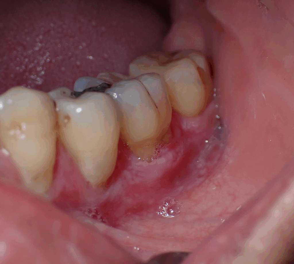
Figure 1:Picture of patient with Oral Lichenoid reaction
Angioedema, a Type I hypersensitivity, manifests as rapid, painless swelling of the lips, tongue, or buccal mucosa. This may progress to airway compromise and constitutes a true dental emergency [8].
Severe drug-induced reactions such as erythema multiforme (EM) (Figure 2), Stevens–Johnson syndrome (SJS), and toxic epidermal necrolysis (TEN) often begin with oral lesions, painful ulcerations, crusted lips, and target-like skin lesions. Prompt drug withdrawal and systemic corticosteroids are critical [9].
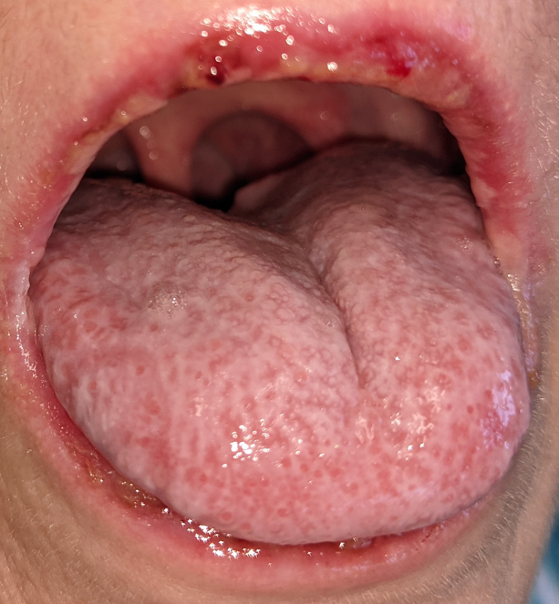
Figure 2. Picture of patient with Erythema Multiforme
Less severe yet clinically significant forms, such as plasma-cell gingivitis (Figure 3) and oral allergy syndrome (OAS), are usually reversible once the allergen, often a flavoring or food protein, is identified and removed [10].
Figure 3. Picture of patient with biopsy proven Plasma Cell Gingivitis
Diagnosis: From Suspicion to Confirmation
Diagnosing hypersensitivity requires detailed history-taking and careful observation.
- Medical and drug history: Correlate symptom onset with new medications, dental materials, or oral hygiene products.
- Clinical features: Note the distribution and morphology of lesions and any systemic symptoms such as fever or malaise.
- Drug withdrawal: Temporarily discontinuing the suspected agent (in collaboration with the prescribing physician) often confirms the diagnosis [1].
- Biopsy and direct immunofluorescence (DIF): When autoimmune mucosal disease (e.g., pemphigus, mucous membrane pemphigoid) is suspected, DIF provides diagnostic clarity [11].
- Patch testing: Referral for patch or allergy testing may be warranted to identify sensitivities to dental materials or topical agents [12].
Interprofessional communication between dentist, physician, and specialist ensures accurate diagnosis and comprehensive care.
Management: Practical Steps in the Dental Setting
Treatment begins by eliminating the offending agent and managing the inflammatory response.
- Remove or replace the trigger. Substitute medications or materials with lower allergenic potential whenever possible [1].
- Topical corticosteroids, such as fluocinonide gel or dexamethasone rinse—reduce local inflammation [1].
- Systemic corticosteroids are indicated for extensive or severe reactions, including EM and SJS [9].
- Antihistamines can alleviate swelling or itching in acute allergic reactions.
- Immunosuppressive agents (e.g., hydroxychloroquine, mycophenolate mofetil) may be required for chronic, steroid-refractory mucositis [11].
- Emergency preparedness: For anaphylaxis, administer epinephrine 0.3 mg IM (1:1000) immediately and activate EMS [8].
- Follow-up: Schedule three-month recall visits to monitor lesion healing and prevent recurrence [1].
Successful outcomes depend on interdisciplinary collaboration among dentists, dermatologists, allergists, and rheumatologists [1].
Clinical Takeaways
- Review and update allergy and medication histories at each visit.
- Recognize that oral lesions may signal systemic hypersensitivity.
- Maintain emergency protocols for allergic reactions.
- Educate patients about early reporting of oral burning, swelling, or ulcerations after medication or restorative changes.
Integrating these practices into daily clinical routines enhances patient safety and supports early identification of immune-mediated disorders.
Conclusion
Hypersensitivity reactions to medications remind us that oral health and systemic immunity are inseparably linked. Whether a mild contact stomatitis or a severe mucocutaneous eruption, these immune events demand careful evaluation and prompt, coordinated care. By staying informed and vigilant, dental practitioners protect their patients from avoidable harm and uphold the highest standards of comprehensive care.
Earn an Online Postgraduate Degree in Orofacial Pain and Oral Medicine
Are you interested in a variety of issues focused on orofacial pain, medicine and sleep disorders? Consider enrolling in the Herman Ostrow School of Dentistry of USC’s online, competency-based certificate or master’s program in Orofacial Pain and Oral Medicine.
References
- Ostrow School of Dentistry of USC. OFPM #726 Immunology & Oral Diseases: Hypersensitivity and VLE Manual. Los Angeles, CA: University of Southern California; 2023.
- Gell PGH, Coombs RRA. Clinical Aspects of Immunology. Oxford: Blackwell Scientific; 1963.
- Khan DA. Allergic and immunologic reactions to drugs and biologic agents. J Allergy Clin Immunol. 2019;143(2):327–338.
- Pichler WJ. Drug hypersensitivity: classification and clinical features. J Allergy Clin Immunol. 2017;140(2):333–349.
- Abbas AK, Lichtman AH, Pillai S. Cellular & Molecular Immunology. 10th ed. Philadelphia: Elsevier; 2022.
- Saini SS. Delayed-type hypersensitivity reactions in clinical practice. Curr Allergy Asthma Rep. 2018;18(11):60.
- Reinhart JP, Stoopler ET, Crawford GH. Oral hypersensitivity reactions: review of clinical presentation, patch testing, and dental material implications. Dermatol Clin. 2020;38(4):467–476.
- Chiu AG et al. Acute allergic angioedema of the lips. Case Rep Otolaryngol. 2016;2016:4872587.
- Roujeau JC, Stern RS. Severe adverse cutaneous reactions to drugs. N Engl J Med. 1994;331(19):1272–1285.
- Ortolani C et al. Oral allergy syndrome. Allergy. 2019;74(10):1960–1972.
- Chan LS. Pemphigus and pemphigoid: diagnosis and management. Clin Rev Allergy Immunol. 2019;56(1):10–25.
- Czerwinski M et al. Patch testing in dental material hypersensitivity. Contact Dermatitis. 2019;81(1):1–10.