X-ray image showing articular fossa and condyle.For TMJ mobilization procedures read: Closed Lock Mobilization: TMJ Exercises & Stretches.
Jaw Range of Motion Assessment
Prior to the procedure, introduce yourself to the patient, explain the purpose of the examination, obtain consent, and be sure to meet infectious control standards.
1. Interincisal Open Measurement
There are three interincisal measurements to assess a patient’s jaw range of motion: pain-free, unassisted, and assisted.
Pain-free
First, you’ll want to see how wide patients can open their mouth without any pain. Ask your patient to “open their mouth as wide as possible without any pain and stop and hold when pain first becomes evident.”
Next, measure the active opening distance by placing your ruler, such as a TheraBite Jaw ROM Scale, on the incisal edge of the mandibular central incisor and measuring the distance to the incisal edge of the opposing maxillary central incisor.
Unassisted & Assisted
Next, measure the total voluntary movement ability of their jaw even if pain is present. For this measurement, ask the patient to “open as wide as possible even if it hurts and hold this position,” and using your ruler, measure the active opening distance as described above.
For the passive or “assisted” measurement, repeat the same process, but this time have your patient open as wide as possible while you apply a small amount of prying pressure with your fingers on their incisor teeth.
Like what you’re learning? Download a brochure for our Orofacial Pain and Oral Medicine certificate or master’s degree program.
2. Lateral Jaw Motion Assessment
Instruct your patient to move their jaw fully to one side with the teeth only slightly apart, and hold that position while you measure the distance. Then do the same movement to the opposite side.
To find the maximum lateral jaw motion distance, measure from the upper centrals where the mandibular central midline is located with the teeth fully together to the mandibular midline when the jaw is fully positioned to either the right or left.
3. Protrusive Jaw Motion Assessment
Lastly, instruct the patient to separate their teeth slightly and move their jaw as far to the right as possible, forward, then hold it while you measure. To measure the protrusive jaw motion, first measure the degree of overjet (labial surface of maxillary central to the labial surface of mandibular central with the teeth fully together), and then with the jaw fully forward, measure the distance from labial surface to labial surface and add the two measurements together.
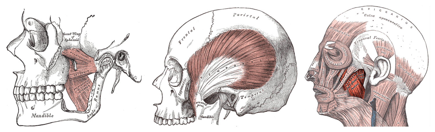
Supplemental Activity: What is the function of the Temporalis muscle in jaw movements?
TMJ Noise and Tenderness Exam
1. Dorsal TMJ Capsule Palpation
Have the patient open their mouth to find the lateral pole of condyle and then move your finger to the back and slightly inferior to the condyle pole to evaluate the dorsal surface of TMJ capsule. Next, have the patient close their mouth and insert your smallest finger into the external auditory meatus. Apply pressure forward to the back of the condyle making sure not to compress the tragus.
2. Lateral TMJ Capsule Palpation
Palpate the lateral capsule with the index finger placed just in front of the tragus of the ear. Ask the patient to open slightly, while you feel for the lateral pole of the condyle. Next, repeat the process by palpitating the lateral TMJ capsule with the mouth closed.
3. TMJ Noise Palpation
Position your middle and index fingers over the lateral pole and slightly in front of the condyle. Instruct the patient to open and close their mouth during the examination, and using very light pressure, listen carefully for audible TMJ noises.
4. TMJ Noise Auscultation Under Loading Conditions
Place a stethoscope over the anterior zygomatic bone, not over the condyle, and listen for joint noise. Next, test under loading conditions. Use one hand to hold the stethoscope on the zygoma in front of condyle, place your other hand under the mandible, and use your fingers to find and apply upward pressure on the inferior bony border of the mandible near the angle. Once you’re in position, instruct the patient to open wide, close, move forward, right, and left with their teeth apart as you listen for joint sounds.
Additional Reading on Orofacial Movement Disorders
Want to learn more about orofacial movement disorders? Check out these other articles by USC Ostrow professors:
Postgraduate Orofacial Pain and Oral Medicine Master’s Degree
Learn more about TMJ disorders by enrolling in our online, competency-based certificate or master’s program in Orofacial Pain and Oral Medicine.